
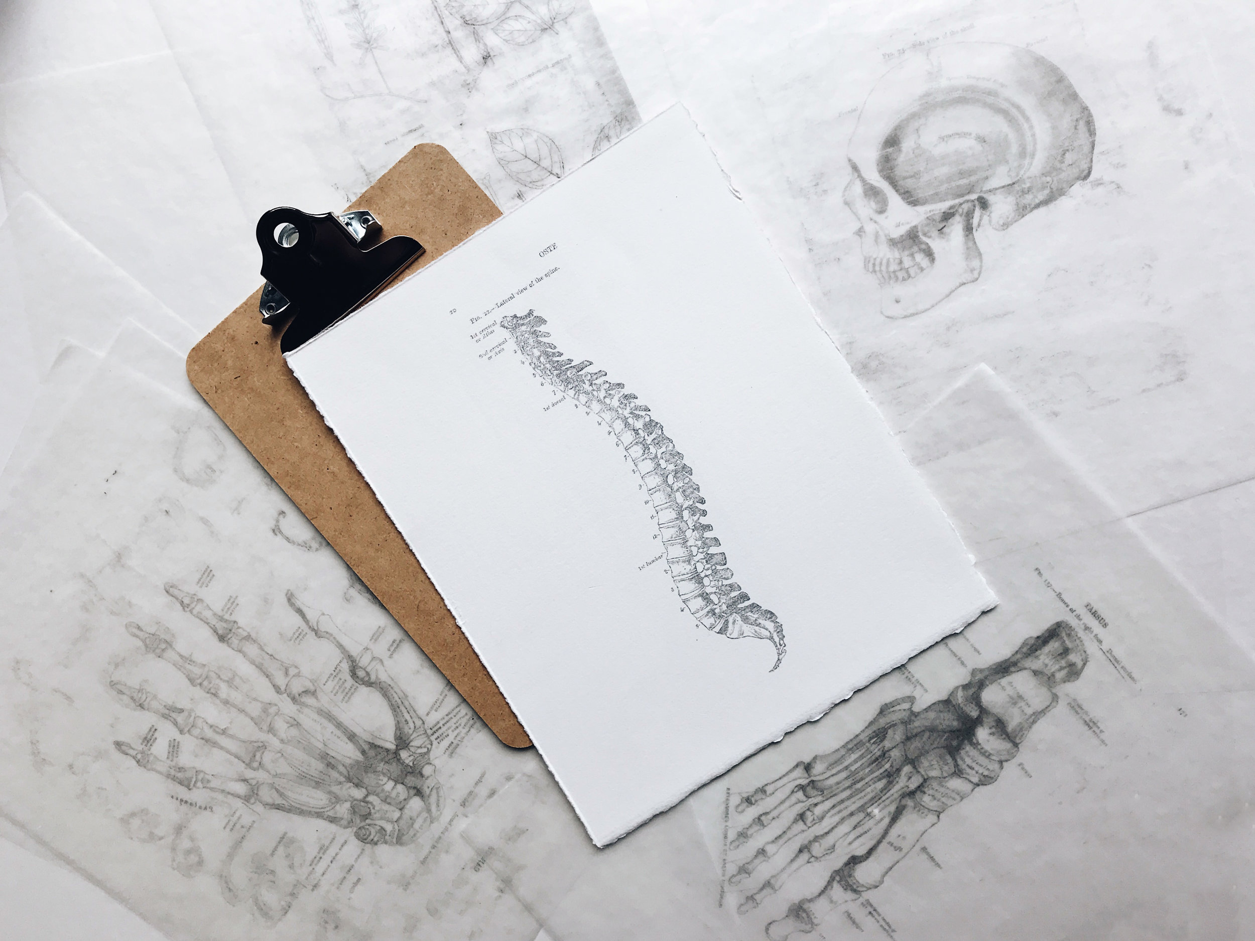
Three things that every person with back pain should be working on.
There are three things that every person with back pain should be working on. General patterns emerge with almost every single person who is experiencing back pain, regardless of what is causing their pain.
If you have back pain there are three things that you should be working on right now to relieve your pain.
I know there are a number of causes for back pain. But there are general patterns that emerge with almost ever single person we see who is experiencing back pain, regardless of what is causing their pain.
The three most common contributing factors that almost all patients with back pain have are:
1.) Poor hip mobility
2.) Poor core muscle strength
3.) Altered breathing patterns
How poor hip mobility leads to back pain:
Hip mobility is key to the success or failure for a majority of the activities that you do. Good hip mobility will allow you to squat down to pick something up while maintaining good spinal posture.
Good hip mobility will also allow you to efficiently walk or run without overextending your spine to compensate.
There are three key directions that your hips need to be mobile into in order to effectively take stress off your low back.

Cara in Supta Badda Konasana pose
1.) Adduction: Being flexible into adduction will allow your pelvis to clear your hips when you squat down to pick something up. By clearing the pelvis through the hips you will be able to squat deeper while maintaining a nice neutral spine. This way you won’t have to round your back so much to when squatting down.
A great stretch to improve adduction is supta badda konasana (yes it’s a yoga pose, you could probably tell by the name) shown here.

Cara demonstrating the pigeon pose.
2.) Flexion: Being able to flex your hip higher will allow you to squat deeper while maintaining your back in a neutral alignment. In order to do this you need to have flexible glutes.
A great stretch to improve hip flexion and glute flexibility is the pigeon pose, shown here.

Cara demonstrating a kneeling hip extension stretch
3.) Extension: Having good hip extension is critical to maintaining a happy and healthy spine when you walk or run. Naturally you need your hips to extend to move through your full gait cycle. If your hips are tight into extension then the body will compensate by extending more into your low back. Over time this could cause irritation and pain in the low back.
A great stretch to improve hip extension is a kneeling hip extension stretch, shown here:
How poor core muscle strength leads to back pain:
Honestly this one is pretty self explanatory. If your core is weak you won’t be able to stabilize your spine when you perform daily tasks. This can easily lead to back pain through overuse. Traditionally this means that your paraspinals (the muscles that run along the length of your spine) will be overworked leading to fatigue and soreness.
Watch this video below to get a better idea of how to properly contract your core:
Now that you know how to properly contract your core there are a number of exercises you can do to strengthen.
How altered breathing patterns lead to back pain:
This one is not so obvious compared to the first two. But how you breathe is critical to stabilizing your back during activities throughout the day.
The diaphragm is one of your core muscles. Possibly one of the most important and least talked about core muscle.
Timing diaphragm breathing to specific movements like squatting and lifting can significantly improve your core stability and reduce strain to the low back.
For example: As you squat down to pick something off the floor you should be taking a nice deep breath in through the diaphragm. The contraction of the diaphragm will increase the intraabdominal pressure making your back more stiff and rigid to lift.
For more info on diaphragmatic breathing I wrote a great article recently that you can check out here: https://www.gofit-pt.com/blog/diaphragmatic-breathing.
In Conclusion:
Back pain can have a number of different causes. However there tends to be a trend among all patient’s with back pain regardless of the cause of the pain. These patient’s all seem to have three things in common. First, they have poor hip mobility. Second, they have poor core muscle strength. Lastly, they do not know how to breath properly and use their breath to stabilize their spine.
To learn how we can help, or to schedule an appointment, click here:

The one thing you should know about pain that no medical professional is telling you.
There is one thing that you should know about pain that nobody in the medical field has told you. Knowing this could completely change how you feel pain.
There are many misconceptions perpetuated by the medical community these days.
One of the most impactful misconceptions that we experience on a regular basis in our physical therapy practice is the misconception about pain.
after reading this article you will have a new understanding of pain.
If a medical professional has ever told you that because you have a bulging disc, or rotator cuff tear, or osteoarthritis, or any other diagnosis, that you will have pain for the rest of your life, this article is for you.
The standard way that pain is taught is that Pain = Tissue Damage and that Tissue Damage = Pain. This idea has been disproven time and time again by research but somehow this misconception lives on.
Ever find a bruise on your body and wonder how it got there? That bruise is a clear sign of tissue damage but somehow it didn’t hurt. How does that happen?
Did you know that there are a number of studies (referenced below) that show a high rate of “abnormalities” (that include disc bulges, herniations, labral tears, rotator cuff tears) in ASYMPTOMATIC (meaning they don’t have pain) individuals?
Did you know that there are likely a high number of people walking around with full blown rotator cuff tears that have no pain or limitation?
How does that happen?
Let me expand on this:
Did you know that there are no nerves in your body specifically designed to sense pain? There are nerves that sense touch, pressure, temperature, and chemicals to name a few. But it is entirely up to your brain on how to interpret those sensations.
Take this example:
Your eyes sense light, it’s your brain that interprets the light based on your experiences and the context of events and then produces an image that we call vision.
Another example is your ears:
The nerves in your ears sense vibration. It is once again your brain that interprets these vibrations based on experience and context and produces what we call sound.
So how does pain really work?
You experience pain when your body perceives a threat.
Let’s say your walking barefoot and you step on a piece of glass. The nerves in your foot sense something sharp and send a signal to your brain. Your brain then interprets that signal and sets off an alarm. That alarm is pain. The brain has decided that the piece of glass is a threat and it makes you experience pain so that you will stop and remove the piece of glass in your foot.
This makes sense right?
Now, lets make this example a little more complicated:
Let’s say you’re awakened in the middle of the night to a ragging fire in your room. What do you do? You run out of the room. But as your running you step on that same piece of glass. What happens now, do you think you feel pain? Definitely not! You stepping on a piece of glass is much less of a threat than the ragging fire in your room. So you run out of the house with the piece of glass in your foot and only until you have safely removed yourself from the threat of fire, do you start to notice that your foot hurts. This is because the bigger threat has been removed and your body can now worry about the smaller one.
So what does this have to do with physical therapy?
In the example above, the brain activates an alarm based on the brains perception of threat. That alarm makes you sense pain. The alarm can be turned on or off depending on the circumstance, seen in the second example.
Once the threat is removed (i.e. once you remove the piece of glass) the alarm starts to turn off and in few minutes the pain is relatively minor. In a few days the pain is gone.
However, what happens when the alarm stays on?
This can happen in about 25% of people that experience pain.
Remember pain is triggered by the body’s perception of threat. The real key here is that THREAT is multifactorial.
Let’s compare the following two examples:
Example 1: Lets say you’re a quarterback for a professional football team and you have injured your throwing shoulder. You’re midway through the season in the last year of your contract, the season hasn’t been going well and your worried that if you don’t perform this could be the end of your career. You’re married with two children who fully rely on the income that you make. You love playing football, you have worked incredibly hard to make it to the professional level and it’s the only thing that you have ever wanted to do in your life.
Example 2: Lets say you were convinced by a friend to play a game of pick up football and you sustain the same exact injury to your throwing arm. You don’t really love playing football, you barely ever play and you have a secure job that doesn’t require more out of your arm than typing on the computer.
Of these two examples who is threatened more by the same injury? Clearly it’s the professional quarterback. Because the repercussions of the injury have more of a potential impact on all aspects of the quarterback’s life. For this hypothetical quarterback this injury could mean that he gets replaced. If he gets replaced then maybe his contract won’t get picked up again. He could loose his job, his source of income, his identity, and even his opportunity to do the one thing that he loves to do, play football. Think his alarm is going off? Absolutely, there are a number of underlying factors that are making his alarm go off like crazy.
Because of these extra factors it is possible that the quarterback’s alarm will keep going off even as the tissues heal.
The quarterback wouldn’t be alone in this. Remember this happens in 25% of people who experience pain and most people don’t even know it’s happening because all they have ever been told is that pain = tissue injury.
Tissues heal, but sometimes the alarm stays on.
Part of a good physical therapy approach is to identify if someone’s alarm is still on.
Treating the sensitive alarm so that it calms down is just as important as treating the tissues.
If you have gone through failed treatment after failed treatment it’s almost a certainty that your alarm is way too sensitive.
There are plenty of ways to turn down the intensity of that alarm. The first and most important is through knowledge. Studies show that just by understanding how pain really works you can start to turn down that sensitive alarm.
Call/Text today To Book an Appointment.
we can help you turn off the alarm!
References:
Jensen MC, Brant-Zawadzki MN, Obuchowski N, Modic MT, Malkasian D, Ross JS. Magnetic resonance imaging of the lumbar spine in people without back pain. N Engl J Med. 1994 Jul 14;331(2):69-73. doi: 10.1056/NEJM199407143310201. PMID: 8208267.
Bastian SA, Rahmi H, Crues J, Bhanu S, Blout C, Rangarajan R, Lee B, Itamura J. Variations of magnetic resonance imaging findings in asymptomatic elbows. J Shoulder Elbow Surg. 2019 Jun;28(6S):S154-S160. doi: 10.1016/j.jse.2019.05.006. PMID: 31196510.
Gutierrez NM, Granville C, Kaplan L, Baraga M, Jose J. Elbow MRI Findings Do Not Correlate With Future Placement on the Disabled List in Asymptomatic Professional Baseball Pitchers. Sports Health. 2017 May/Jun;9(3):222-229. doi: 10.1177/1941738117701769. Epub 2017 Apr 10. PMID: 28394713; PMCID: PMC5435154.
Gill TK, Shanahan EM, Allison D, Alcorn D, Hill CL. Prevalence of abnormalities on shoulder MRI in symptomatic and asymptomatic older adults. Int J Rheum Dis. 2014 Nov;17(8):863-71. doi: 10.1111/1756-185X.12476. Epub 2014 Oct 8. PMID: 25294682.
Hacken B, Onks C, Flemming D, Mosher T, Silvis M, Black K, Stuck D, Dhawan A. Prevalence of MRI Shoulder Abnormalities in Asymptomatic Professional and Collegiate Ice Hockey Athletes. Orthop J Sports Med. 2019 Oct 10;7(10):2325967119876865. doi: 10.1177/2325967119876865. PMID: 31637270; PMCID: PMC6787880.
Louw A, Zimney K, Puentedura EJ, Diener I. The efficacy of pain neuroscience education on musculoskeletal pain: A systematic review of the literature. Physiother Theory Pract. 2016 Jul;32(5):332-55. doi: 10.1080/09593985.2016.1194646. Epub 2016 Jun 28. PMID: 27351541.

Overuse injuries explained.
Learn what overuse injuries are, why they occur, and what you can do to fix them.
Learn what overuse injuries are, why they occur, and what you can do to fix them.
What is an overuse injury?
An overuse injury is pretty much exactly how it sounds. You ended up overloading an area of your body through a particular activity or combination of activities and this has resulted in injury.
This can happen for a number of reasons but often times the cause is more complex and possibly multifactorial.
Why did I develop an overuse injury in my leg?
To answer this question let’s dust off that old high school physics book (if you still have it and let’s face it, you don’t). You may remember being taught that for every action there is an equal and opposite reaction.
This is absolutely true when we walk, jog, or sprint. When we take a step, we are exerting a force on the ground. The ground is also exerting a force back through us (equal and opposite). This is termed the ground reaction force.
A changing ground reaction force:
Whenever you take a step, or even stand still for that matter, the ground is exerting a force through your body.
The force is constantly changing. For instance, the ground rection force gets larger with more intense activity. Stand still and the force is relatively small, walk and it goes up a little bit, jog it goes up a little bit more, sprint and it goes up even further.
The type of surface we walk or run on makes a difference too. Harder surfaces will produce more ground reaction force while softer ones will produce less. Think about running on cement vs. running on a dirt trail. I’m sure you noticed that the trail feels softer and easier.
The type of shoe (or lack thereof) makes a difference. Shoes with more cushion will reduce the ground reaction force just as running barefoot will increase it.
So why not just wear super cushiony shoes and run in mud?
There is always a catch and here it is: with all things being equal a softer surface will make you slower and a harder surface will make you faster. A shoe with more cushion will make you slower than running barefoot. Think about sprinters, they don’t wear big cushiony trainers, they wear slim track spikes.
So, what does all this have to do with injury?
Thanks for hanging in there with me.
This could start to explain why you may have noticed pain after a particular run. Maybe you ran a different loop and the surface was harder. Maybe your shoes having been wearing out and they are no longer giving you that little bit of extra shock absorption that you needed.
These are a couple of the possible external variables that could have sparked an overuse injury.
There are however some intrinsic reasons why this happened as well.
Your body must be able to control the ground reaction force
This is where a lot of problems begin. Back to physics…
Remember your body needs to be able to meet the ground reaction force when you move. In a perfectly balanced, mobile, and strong body the force will be equally taken up and distributed throughout the joints, muscles, and tendons.
But what happens if, let’s say, your hip is a little weak?
Well in that case something else along the path will have to take up the slack. This could be around the knee or the ankle for starters.
Let’s just say for the sake of examples your quad is the muscle that makes up for your weak hip…. Do you see where this is going? Can you say runner’s knee(chondromalacia patella) or patellar tendinopathy?
Or maybe it’s your foot or ankle. Hello plantarfascitis, Achilles tendinopathy, or tibialis posterior syndrome.
Typically, it tends to be the large muscles that aren’t pulling their fair share of the load and the poor little muscles get beat up as a result.
You can fix it.
Even though the problem likely started without you even knowing, there are ways that you can fix it.
Here are 3 tips to get you back on track:
1.) Cut down the intensity of your activity to a level that is non-aggravating. There isn’t necessarily a reason to stop altogether. You just have to bring it back down to a level that your body can handle. This likely means cutting the intensity and/or duration of your exercise.
2.) Get stronger in your core, glutes, and quads. Add two to three glute and quad strengthening activities 3 times a week to your routine.
3.) Build back slowly. The safest return to activity is at an increase of 5-10% (either intensity or duration) every week. If you have time be conservative and progress slower at a 5% rate, do it, your body will thank you.

One exercise to improve core strength, reduce stress, and improve muscle recovery.
One exercise to improve core strength, reduce stress, and improve muscle recovery: diaphragmatic breathing.
Is there really one exercise that can do it all?
Yes, there is. And, it won’t even make you sweat!
This blog post is a follow up to a recent Facebook and Insta post.
Diaphragmatic breathing can have a huge impact on many aspects related to our health.
For instance, did you know that diaphragmatic breathing exercises can actually improve your balance?!? Whaaaaat?
I know, it sounds crazy, but it’s true. Let’s take a closer look at how the diaphragm can improve your core health:
How the diaphragm can improve your core stability:
We typically use the concept of a cylinder to explain how the core works.
If we think of the core as a cylinder the diaphragm is on top, the pelvic floor on the bottom, and a number of muscles including the transversus abdominus make up the sides of the cylinder.
The diaphragm’s role is to regulate the amount of intra-abdominal pressure.
Try this: Contract your core and then try to take a slow breath in through your nose. Your core gets tighter, doesn’t it?
That’s the diaphragm doing its job to increase the pressure within your core. This helps stiffen the spine and hold it in position.
This concept is backed by scientific evidence that demonstrates improvement in core strength with diaphragmatic breathing exercise. (5)
It is no surprise then that poor breathing patterns have been linked to neck pain, thoracic, pain, low back pain, and even balance dysfunctions. (4)
If your core isn’t functioning properly, how can one expect to stay pain free?
It should also be no surprise that there are studies that show that regular diaphragmatic breathing can improve pain and balance. (1,4)
Diaphragmatic breathing to reduce stress.
Diaphragmatic breathing has been basking in its stress reduction glory for years. Buddhist monks, à la Tich Nhat Hanh, have been preaching about the effects of slow deep diaphragmatic breathing on stress reduction, mindfulness, and peace for decades.
But how does it work?
The answer lies with the Vagus nerve.
The Vagus nerve innervates the diaphragm, along with a number of other organs throughout the body.
The Vagus nerve is intimately linked with the parasympathetic nervous system.
The parasympathetic nervous system is responsible for rest and recovery. When the parasympathetic nervous system gets activated heart rate slows down, good endorphins are released, and capillaries dilate. This allows for our bodies to relax and recover. (3)
When we breathe with our diaphragm, we stimulate our Vagus nerve. Specifically diaphragmatic breathing massages our Vagus nerve as it passes near and through the diaphragm. This sort of massage to the Vagus nerve can only occur if our diaphragm is contracting and relaxing. When the Vagus nerve is stimulated in this way it sends signals throughout the body to do all the wonderful things that we relate to the parasympathetic nervous system. (2)
So now we know that diaphragmatic breathing can activate the parasympathetic nervous system and improve core stability.
What else can it do?
Diaphragmatic breathing to improve muscle recovery
Diaphragmatic breathing normalizes blood chemistry and improves blood flow and oxygen to your muscles. (7)
Muscles need good blood flow, oxygen, and balanced blood chemistry to heal properly.
Regular diaphragmatic breathing can be a simple daily exercise that you do to promote good muscle recovery.
Excited to try it? Here is how to get started:
The diaphragm is a muscle, a strong muscle, and you can consciously control how quickly, slowly, or forcefully it contracts.
First, envision you have a belt or rubber band that wraps around the lower part of your rib cage.
Next, take a slow deep breath in through your nose and try to expand out against your imaginary belt or rubber band in 360 degrees.
Then slowly exhale through your nose
If done correctly you will feel like you are pulling into your lungs through your rib cage rather than sniffing it in through your nose! A well controlled diaphragmatic breath should be relatively quiet.
Work over the next couple minutes to lengthen the breath in and out to around 3-5 seconds. Over time you can work to increase this length to upwards of 7 seconds.
As you do this over the next couple minutes feel your heart rate lower and your mind getting calm. Amazing right?
Start by adding a couple minutes worth of diaphragmatic breathing in throughout your periods of rest each day.
Then begin to be more conscious of your breath throughout longer portions of the day. Before you know it, you will be a true diaphragm breather (and probably a lot calmer!).
Other amazing benefits of diaphragmatic breathing:
Did you know that diaphragmatic breathing can reduce the effects of motion sickness(8).
Diaphragmatic breathing has been showing to reduce the symptoms of irritable bowel (IBS) and GERD(6).
Want more? check out this vid below:
References:
1. Anderson BE, Bliven KCH. The Use of Breathing Exercises in the Treatment of Chronic, Nonspecific Low Back Pain. J Sport Rehabil. 2017;26(5):452-458. doi:10.1123/jsr.2015-0199
2. Bordoni B, Morabito B. Symptomatology Correlations Between the Diaphragm and Irritable Bowel Syndrome. Cureus. 2018;10(7):e3036. Published 2018 Jul 23. doi:10.7759/cureus.3036
3. Bordoni B, Purgol S, Bizzarri A, Modica M, Morabito B. The Influence of Breathing on the Central Nervous System. Cureus. 2018;10(6):e2724. Published 2018 Jun 1. doi:10.7759/cureus.2724
4. Bradley H, Esformes J. Breathing pattern disorders and functional movement. Int J Sports Phys Ther. 2014;9(1):28-39.
5. Cavaggioni L, Ongaro L, Zannin E, Iaia FM, Alberti G. Effects of different core exercises on respiratory parameters and abdominal strength. J Phys Ther Sci. 2015;27(10):3249-3253. doi:10.1589/jpts.27.3249
6. Eherer AJ, Netolitzky F, Högenauer C, et al. Positive effect of abdominal breathing exercise on gastroesophageal reflux disease: a randomized, controlled study. Am J Gastroenterol. 2012;107(3):372-378. doi:10.1038/ajg.2011.420
7. Schleifer LM, Ley R, Spalding TW. A hyperventilation theory of job stress and musculoskeletal disorders. Am J Ind Med. 2002;41(5):420-432. doi:10.1002/ajim.10061
8. Stromberg SE, Russell ME, Carlson CR. Diaphragmatic breathing and its effectiveness for the management of motion sickness. Aerosp Med Hum Perform. 2015;86(5):452-457. doi:10.3357/AMHP.4152.2015
Is your headache from neck pain or your neck pain from a headache?
Headaches and neck pain are intimately linked. Find out more here.
How often do people experience neck pain?
Neck pain is very common in the U.S. with some estimates suggesting that 70% of the population will experience neck pain at some point. Though not as common, 1 in 6 Americans are likely to suffer from severe headache or migraines.
We commonly see clients who suffer from both neck pain and headaches. It turns out that these two seem to go hand in hand.
Are neck pain and headaches linked?
Studies have shown that 90% of people with tension type headaches have neck pain, 75% of people with migraines have neck pain, and 84% of people with self reported sinus headaches have neck pain(1,2,3).
So what is causing what?
Is the headache coming from the neck pain or is the neck pain being driven by the headache? The answer; it depends.
There have been numerous studies, based on the work of Janet Travel, that have looked at myofascial (muscular) origins of headaches.
Similarly, there is research that links joint mobility issues in upper part of the neck to headache symptoms(4).
It’s not so easy, especially in a blog post, to be definitive of the origin of every headache or neck pain.
The best bet is to have a skilled physical therapist perform a thorough evaluation to look for all possible contributing factors to your headaches.
Conclusion:
The take home message is this: Your neck pain or headache, or both, is treatable! If you thought that there was nothing else you could do for your headache or neck pain besides rest and medication, you were wrong.
Muscles and joints can both be common causes of neck pain and headaches. Both sources of pain are treatable. It will take a skilled clinician to determine the source, or sources, of the pain and treat them accordingly.
References:
1. Ashina, S., Bendtsen, L., Lyngberg, A. C., Lipton, R. B., Hajiyeva, N., & Jensen, R. (2015). Prevalence of neck pain in migraine and tension-type headache: A population study. Cephalalgia, 35(3), 211–219. https://doi.org/10.1177/0333102414535110
2. Burch, R., Rizzoli, P., & Loder, E. (2018). The prevalence and impact of migraine and severe headach in the united states: Figures and trends from government health studies. Headache, 58(4), 496-505.
3. Malo-Urries, M., Tricas-Moreno, JM., Estebanez-de-Miguel, E. et al. (2017). Immediate effects of upper cervical translatoric mobilization on cervical mobility and pressure pain threshold I patients with cervicogenic headache: A randomized controlled trial. Journal of Manipulative and Physiological Therapeutics, 40(9), 649-658.
4. Shannon M. Petersen, Gwendolen A. Jull & Kenneth E. Learman (2019) Self-reported sinus headaches are associated with neck pain and cervical musculoskeletal dysfunction: a preliminary observational case control study, Journal of Manual & Manipulative Therapy, DOI: 10.1080/10669817.2019.1572987
Improving your core muscle coordination to reduce back pain
Find out how improving your core muscle strength and coordination can reduce low back pain.
Back pain is one of the leading reasons why people seek physical therapy care. GOfit is no exception as we see many patients who experience back pain.
There can be numerous causes of back pain from muscles to joints to discs.
What happens to the core muscles in response to pain?
Regardless of the source of pain, there seems to be a similar response of the deep core stabilizer in the presence of pain; it shuts down.
Well, “shuts down” may be a bit of an exaggeration. It doesn’t stop working altogether, but it does begin to atrophy and lose its coordination or muscle timing.
Plenty of studies have shown that specific exercises can cause core muscles to grow, but only a few have looked at how specific exercise effects the timing, or coordination of these muscles. As they say ‘timing is everything.’
Why do core muscles need to be coordinated?
Pure muscle bulk is great but muscles needed to be coordinated in their activity so they can activate at the right time to support the structure of the back.
A delay in activation of the transversus abdominis (one of the main core muscles) of even a few milliseconds could be the difference between pain free and painful functioning.
Think of doing a heavy deadlift or even lifting your child off the floor. If your legs start to move before your core has engaged there will be a lot of extra stress through the low back. This is the classic “I threw my back out” example.
One study, referenced below, looked at the effect that specific exercise had on the timing of transversus abdominis activation.
Subjects of the study performed three exercises of increasing difficulty (abdominal drawing in maneuver, side planks, and bird dog) two times a week for four weeks.
After four weeks muscle activation timing of the transversus abdominis was retested.
The authors found that muscle timing of the transversus abdominis improved significantly in the experimental group compared to controls.
So, what does this all mean?
First, it means that we can improve the activation timing of one of our deep core stabilizers through some pretty simple exercises.
Muscle timing is an important part of coordination.
Having a coordinated and strong core will undoubtedly lead to reduced pain and improved daily functioning.
Selkow NM, Eck MR, Rivas S. Transversus abdominis activation and timing improves following core stability training: A randomized trial. Int J Sports Phys Ther. 2017;12(7):1048-1056.

Achilles tendinopathy: What is it and how can I get it better?
Achilles tendinopathy can be a serious irritation, especially if the activity you love involves walking, jogging, jumping, or running. This post will teach you all you need to know about the problem and ways to get it better.
In this blog we look to explain the process of Achilles tendinopathy.
We will talk about common risk factors, proven treatments, new treatment ideas, and what doesn’t work.
Achilles tendinopathy is one of the most common ankle overuse injuries and is more likely to be found in active individuals who participate in running and jumping sports.
Achilles tendinopathy can be a frustrating condition because of the potential for pain to become persistent and significantly limit athletic participation and daily function.
Don’t worry though there are actionable steps that you can take to to improve this condition.
But first let’s learn a little more about the problem itself.
Your tendon is not inflamed!:
First it is important to point out that we are using the term “tendinopathy” rather than “tendinitis.”
The term tendinitis had been used for decades but it turns out this is actually a bit of a misnomer. The term tendinitis implies that there is an inflammatory condition occurring.
Recent studies however have found that tendon changes occur as a result of failed healing where inflammation and inflammatory cells are not found.
The tendon itself, like most tendons, has a relatively low blood supply. In the Achilles the area of the tendon that is least vascularized is approximately 2 to 6 cm above its insertion into the heel. This explains why this is the most likely area of the tendon to be injured.
Achilles tendinopathy is actually a stalled healing process:
This section is going to be a bit technical, but bare with me because it is important.
In normal tendon healing an injured tendon must go through the four phases of healing.
The first phase, the inflammatory phase, typically lasts two to three days and is important in laying the ground work for healing to occur.
After two to three days if everything goes according to plan the second phase of healing starts to occur. This second phase is called the proliferative phase. This is where new fibrous like tissue is laid down in the area of injury. This typically occurs for 6 weeks until healing starts to enter its next phase: the remodeling phase.
The third phase of healing is the remodeling phase. This is here new tissue starts to align itself in a direction that best absorbs the load through the tendon. This typically lasts another 4 weeks.
Finally, if all goes well, after approximately 10 weeks from the moment of injury the tendon will enter the final phase of healing, the maturation phase.
In this final phase the new fibrous tissue gradually changes to scar tissue over the course of about a year.
That means that if everything goes right it takes an injured tendon about a year to fully heal!!!
So if everything goes right it takes a full year for an injured tendon to heal. Now, that doesn’t mean that you will be out of commission for that full year. You may have healed up sufficiently enough that you can go back to your sport without any pain or limitation.
Its important to know though that the one year mark is still the length of time it will take for the tendon to go through its full healing process.
Tendinopathy is stalled healing:
A tendinopathy occurs when you don’t make it through all four stages of healing.
In fact you end up getting stuck in the second phase, the proliferative phase, of healing.
Being stuck in the proliferative phase leaves a tendon with too many cells responsible for breaking down the tendon. So you get caught in a position where there is too much breakdown vs. buildup.
The tendon can then become weaker and painful. This doesn’t sound like something anyone wants.
Prevention is always the best medicine:
The first question one might want to ask after reading all of the above is: how can someone avoid getting Achilles tendinopathy?
This may be best answered by looking at a list of risk factors associated with Achilles tendinopathy.
Risk Factors:
The first grouping of risk factors are intrinsic factors associated with this disorder. Intrinsic factors include a flat foot, a foot with too much arch, limited mobility of the subtalar joint (heel), and a leg length discrepancy.
Systemic inflammatory conditions such as rheumatoid arthritis, hypertension, diabetes, obesity, hypertension, and certain medications can all increase the risk of developing Achilles tendinopathy as well.
Extrinsic factors include excessive loading of the tendon through sport, training errors that include abrupt changes in frequency or intensity of training, change in training surface (hills or hard surfaces), poor shock absorption and uneven footwear may all contribute to Achilles tendinopathy.
There are a number of factors on this list that can be modified by training and lifestyle changes. Reducing your overall inflammatory lifestyle through diet could easily reduce the risk of developing Achilles tendinopathy.
Likewise, following a gradual progression of exercise and activity without huge jumps in training can significantly reduce your risk.
The bottom line is there are plenty of risk factors on this list that are within your own control!
Now that we know what Achilles tendinopathy is and how it likely got there lets figure out how to get rid of it.
There are a number of different approaches that one may encounter in the medical community. So, let’s review the effectiveness of those approaches.
What doesn’t work
NSAIDs: There is little scientific basis for the use of non-steroidal anti-inflammatory (NSAIDs) medications. As we know from part 1 of this blog post, Achilles tendinopathy is NOT an inflammatory process. So, medications that work to reduce inflammation would add no value to your ability to heal.
Unless you like unnecessary side effects from medication, stay away from the NSAIDs.
Corticosteriod Injections: Just like NSAIDs cortisone is an anti-inflammatory and has no medical rationale for it’s use on this condition.
If that’s not enough, adverse effects were reported in 82% of corticosteroid trials that included further tendon atrophy, tendon rupture, and decreased tendon strength.
This is a no brainer, cortisone has no business being in your Achilles tendon.
What might work
The jury, aka the scientific community, is still out on these interventions.
Some studies have shown some success but there is still more research that needs to be done before the scientific community puts their seal of approval on these treatments.
Extracoporeal shockwave therapy: This is a treatment technique that uses high energy acoustic waves to deliver a mechanical force to the body’s tissues. The workings of this treatment are poorly understood but there have been recent clinical trials that show good results for those with chronic Achilles tendinopathy.
Unfortunately, there needs to be more high-level evidence and specific treatment parameters identified before this treatment moves up the list.
Platelet-rich Plasma (PRP): PRP is the injection of platelet-rich plasma into a site of tendon injury. PRP stimulates healing through a number of growth factors in the plasma. There is a lot of good evidence out there that PRP can help tendon healing elsewhere in the body. Unfortunately, there is not a lot of evidence specific to the Achilles tendon.
Prolotherapy: Prolotherapy is the injection of hyperosmolar dextrose into the tendon. Clinical trials have demonstrated a reduction in pain, improvement in tendon strength and reduction in the size of intratendinous tears associated with the disorder.
Although early research shows benefits with this procedure there still needs to be more high-level studies performed before this treatment is deemed truly effective.
Low level laser therapy: There is conflicting evidence to suggest low level laser therapy is beneficial to Achilles tendinopathy. There are randomized control trials that demonstrate improvement in tendon healing and elasticity.
However other trials showed no effect on pain compared to controls. There is high level evidence to suggest that low level laser therapy may be an effective treatment but more studies are needed to confirm this.
Ultrasound: In animal studies ulatrsound has been shown to stimulate tissue growth and tendon healing. In the acute phases of tendon injury, it has been shown to reduce pain, swelling, and improve patient function.
However there has not been a lot of new evidence to continue to demonstrate its efficacy and for this reason it stays in the “what might work” section of this post.
What does work
Eccentric Exercise: Eccentric exercise remains the gold standard for treating Achilles tendinopathy. There is good high-level evidence for the benefits of eccentric exercise in the management of Achilles tendinopathy.
The mechanism behind the benefits of eccentric exercise are not well understood. Theoretically eccentric exercise produces rapid strengthening of the calf muscle, stiffening of the tendon, and lengthening of the tendon.
Studies have shown some adverse effects if proper form or insufficient rest between bouts of exercise occur so it is important that a physical therapist prescribe the correct dosage of exercise. Most research recommends a course of 6 to 12 weeks of eccentric exercise.
Deep friction massage and tendon mobilization: Studies have demonstrated that deep friction massage combined with stretching can increase tendon healing, improve elasticity of the tendon, and reduce strain through the muscle-tendon unit.
In Conclusion
Achilles tendinopathy is an overuse injury. Over the course of time too much stress was being put through the Achilles. Because of this an injury occurred and that injury is stalled in the second phase of healing.
All the most effective treatments work to progress the tendon through all four phases of healing.
There are easy actionable steps that you can take today to get your Achilles on the path to recovery.
With the right treatment approach and a gradual, progressive return to your activity there is no reason why you can not be rid of your Achilles pain in the future
References:
Li H & Hua Y. Achilles tendinopathy: current concepts about the basic science and clinical treatments. BioMed Research International. 2016(6). https://dx.doi.org/10.1155/2016/6492597.
Chaudhry, F. Effectiveness of dry needling and high-volume image-guided injection in the management of chronic mid-portion Achilles tendinopathy in adult population: a literature review. Eur J Orthop Surg Traumatol. 2017(27). 441-448.
Factors contributing to prolonged back pain
There are a lot of factors that can contribute to someone having low back pain. Not all of them may be obvious to you though. Read this blog to find out more.
Green BN, Johnson CD, Haldeman S, Griffith E, Clay MB, et al. (2018) A scoping review of biopsychosocial risk factors and co-morbidities for common spinal disorders. PLOS ONE 13(6): e0197987. https://doi.org/10.1371/journal.pone.0197987
This week’s blog post goes a little outside the normal topics covered on this blog. Usually we are concerned with mechanical issues or neuromuscular re-education strategies that we can directly influence and effect as physical therapists. But looking at our client as a whole person, it is important to consider and acknowledge other factors that may be contributing to their pain or functional limitation. These are factors that may be outside the scope of physical therapy practice, but it is important to recognize their potential contribution to our client’s complaints.
The authors of this paper were interested in determining risk factors, co-morbidities, and prognostic factors associated with the most common spinal disorders. As the authors point out spinal disorders contribute a significant burden on society and these problems have risen sharply in the past three decades. Spinal disorders are the leading cause for years lived with disability throughout the entire world. If spinal disorders have been getting worse and worse it certainly begs the questions, Why? What has changed in society that has led to this dramatic increase over the past 30 years. The answer seems to be lifestyle.
The authors looked at a number of the most common spinal disorders including joint pain, myofascial pain, radicular pain, trauma, infection, arthritis, and congenital deformities to determine common risk factors associated with these groups of disorders. The authors found that many of the risk factors associated with spinal disorders are completely modifiable and it all comes back to lifestyle.
Let’s start with the obvious. It is well known that living a sedentary lifestyle and over indulging in poor food can lead to numerous health problems, including spinal disorders. Those who had a higher body mass index and did not participate in regular exercise were at high risk for developing and sustaining spinal pain. What you put in your body, in terms of medication and tobacco, can increase your risk for developing a spinal disorder. But it is not just diet and exercise. Work life can be a risk factor for back pain. A repetitive physically demanding job can increase your risk for back pain just as much as a stressful job or a sedentary job. Work stress and other forms of psychological stress can increase the risk of developing or sustaining back pain. There were too many specific risk factors to cover in this blog but it is important to know that the majority of risk factors described in the research are completely modifiable through lifestyle changes.
The take home message here is that there are psychological, nutritional, and physiological changes that we can all make to help reduce the risk of developing and contributing to back pain. We need to continue to champion healthy lifestyle practices that include regular exercise, proper nutrition, healthy work environments, and reduced psychological stress so that we can live healthy, happy, pain free lives.
Movement retraining for hip impingement part 2
A continuation from our previous post. Find out how different hip shapes can have an affect on pain and function.
Lewis C, Loverro K, Khuu A. Kinematic differences during single-leg step-down between individuals with femoroacetabular impingement syndrome and individuals without hip pain. J Orthop Sports Phys Ther. 2018;48(4). 270-279.
This week’s post is a close continuation of our last blog post. This post will hopefully drive home the message that movement retraining is a significantly important piece of rehabilitation. The focus of this paper is on femoracetabular impingement (FAI). FAI is thought to occur when the femoral head (ball) prematurely contacts the acetabulum (socket). When the ball part of the hip joint prematurely contacts the outer rim of the socket the tissue in between (labrum and cartilage) can become damaged and produce pain that is commonly felt in the groin.
As the authors point out there has been a lot of research on different hip joint shapes(morphology) and the relationship between hip joint morphology and the occurrence of FAI and groin pain. On the other hand, there has not been much research on altered movement patterns that may be associated or contributing to FAI. These authors wanted to change just that.
The purpose of this study was to determine if there were differences in movement patterns in those with FAI and those without. Participants ranged in age from 14 years old to 50 years old. Participants in the FAI group had to be diagnosed via physical examination, clinical presentation, and imaging with some form of CAM, pincer, or mixed hip joint morphology. Individuals in the comparison group had to have no recent history of lower extremity pain and were picked as demographic matches.
3D motion analysis was used to measure various movement angles in the pelvis, hip, and knee while participants were instructed to perform a single leg step down. The authors believed the single leg step down would be challenging enough to identify movement discrepancies between groups of participants compared with other dual leg tasks such as the squat.
The results of the study were not completely surprising. The group with FAI had greater hip flexion and anterior pelvic tilt than the pain free group. Increased hip flexion and anterior pelvic tilt would put someone in a position more likely to experience FAI. This suggests that FAI is not necessarily solely a structural issue of the hip joint and that movement patterns may be a modifiable contributor to their pain. People with FAI may be able to retrain their movement patterns to avoid positions that cause impingement in the hip. The authors also found that women had greater hip flexion, adduction, and anterior pelvic tilt which is not surprising given that women typically have greater pelvic width and are more likely to experience impingement symptoms.
So it is likely that people who are experiencing FAI may have an underlying bony morphology but somewhere along the way they began moving in a way that set them up for impingement symptoms. There could be a number of reasons or causes for this altered movement, but it is important to note that movement patterns are changeable.
Movement retraining as a treatment for hip impingement
Retraining the muscles surrounding your hip can make a huge difference if you are experiencing hip or groin pain.
Harris-Hayes M, Steger-May K, Van Dillen L, Schootman M, Salsich G, Czuppon S et al. Reduced hip adduction is associated with improved function after movement-pattern training in young people with chronic hip joint pain. J Orthop Sports Phys Ther. 2018;48(4). 316-324.
The topics of hip impingement and hip pain are near and dear to our hearts here at GoFit and a clinical interest of ours. The article reviewed here uses the term hip joint pain to encompass a number of potential and often related diagnosis (femoracetabular impingment, developmental dysplasia, labral tears, and chondral lesions).
Hip joint pain is a big problem. Especially for young, athletic women. Unfortunately, there isn't a great amount of high-quality research to suggest the best method of treatment. Despite this there has been a huge growth in the amount of surgeries to treat these various conditions.
We talk a lot about manual therapy on the blog, likely because we are manual therapists and that is where our interests lie. But changing movement patterns is just as important in improving pain and function long term. This article is interesting because it looked at the effect movement retraining would have on individuals with chronic hip joint pain.
The patients who participated in this study were between the ages of 18-40 years old, mostly women, who had deep hip pain or groin pain that had been present for at least 3 months. The pain had to be reproduced with a classic impingement test that brought the hip into flexion, adduction, and internal rotation. The authors used some pretty cool 3D kinematic technology to see how the effects of specific movement retraining would have on the person’s ability to correctly perform a single leg squat. They also looked at hip abductor strength and even looked to see if there were structural changes to the head of the femur w/ MRI imaging.
Participants in the study had 6 one-hour sessions over a 6-week period. During these sessions a physical therapist provided instruction in movement of daily functional tasks and patient specific tasks with practice throughout. The patient was asked to perform the specific movements that they were deemed independent with as their home exercise program.
The authors hypothesized that this specific movement retraining would reduce the amount of relative hip adduction the patient had during a single leg squat. This is important because increased hip adduction has been associated with "impingement'' type pain. They also believed that hip abductor strength would increase over the course of treatment.
This was a relatively complex study in the sense that there were a lot of moving parts. With this study, as with most studies, making generalizations can be difficult and results should be taken cautiously. With that being said, there appears to be a couple of take home messages here. The first is that retraining movement during patient specific and functional tasks did work to reduce patient's hip adduction during specific movements. Those who had a greater reduction in hip adduction also had a greater improvement in pain and function assessed through patient specific scales. Interestingly there was no change in hip abductor strength. This suggests that motor control and the timing of muscle activation may play a more important role in stabilizing the hip than pure strength. Also, of note the patients that reported a greater compliance to their home exercise program showed a greater reduction in hip adduction.